About
Meet Your Orthodontist
Team
Why McDonough?
Our Lifetime Guarantee
Before and After
Office Tour
Dentofacial Orthopedics
Our Videos
Blog
Patients
FAQ
Your First Visit
Payment Options
Two-Phase Treatment
Children’s Orthodontist
Adult Orthodontics
Choosing an Orthodontist
Services
Braces
Types of Braces
Types of Appliances
Bonding
Clear Aligners
Aligners FAQ
Aligners Videos
Retainers
Dental Monitoring
Contact Us
Request Appointment
Murray Office
Refer A Friend
Refer A Patient (for Doctors)
Careers
(801) 266 2662
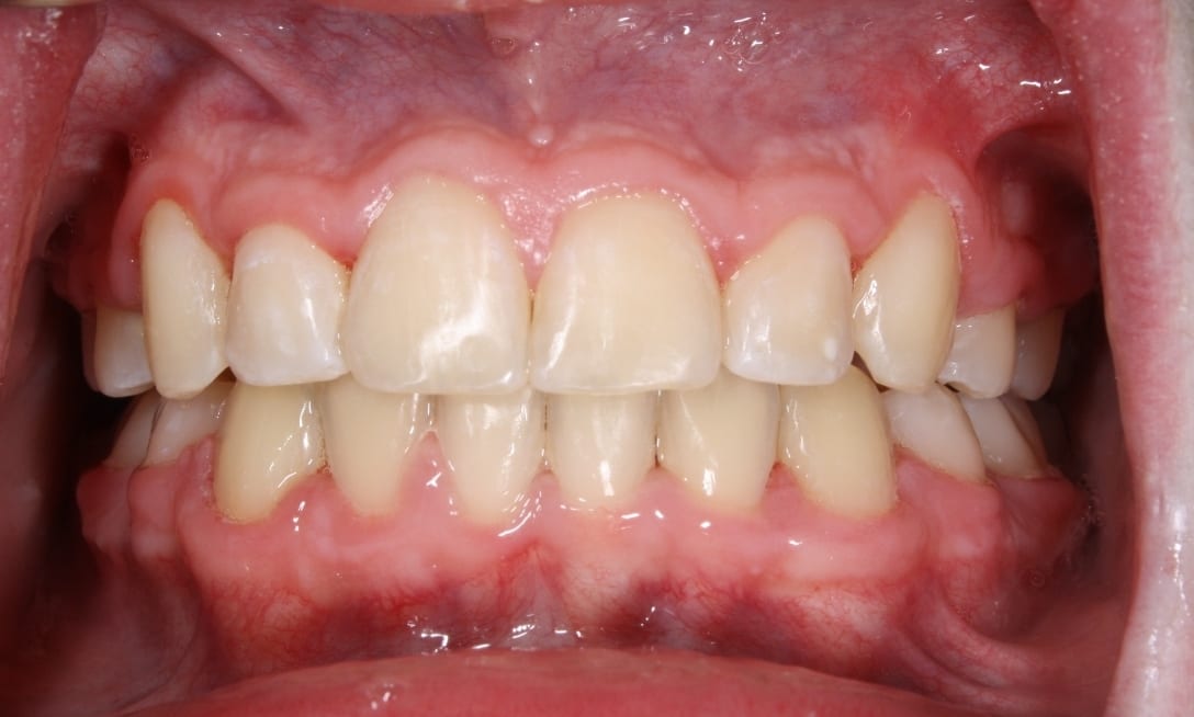
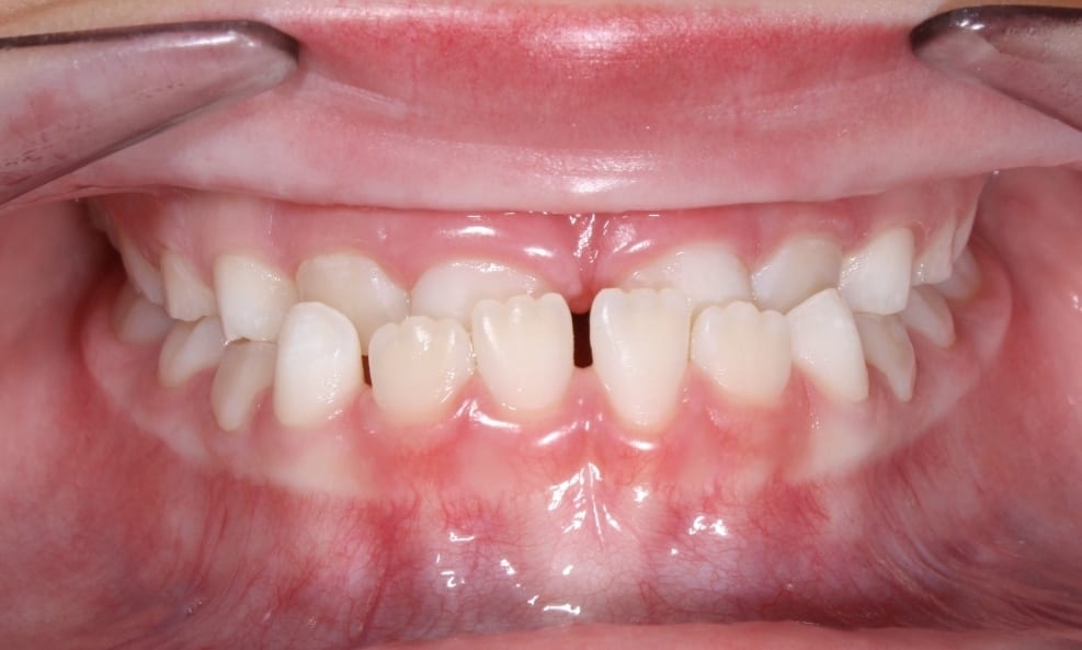
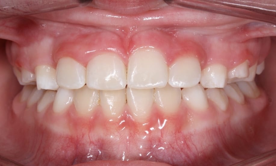
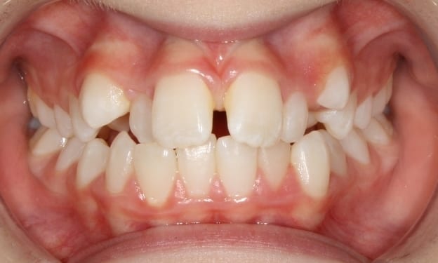
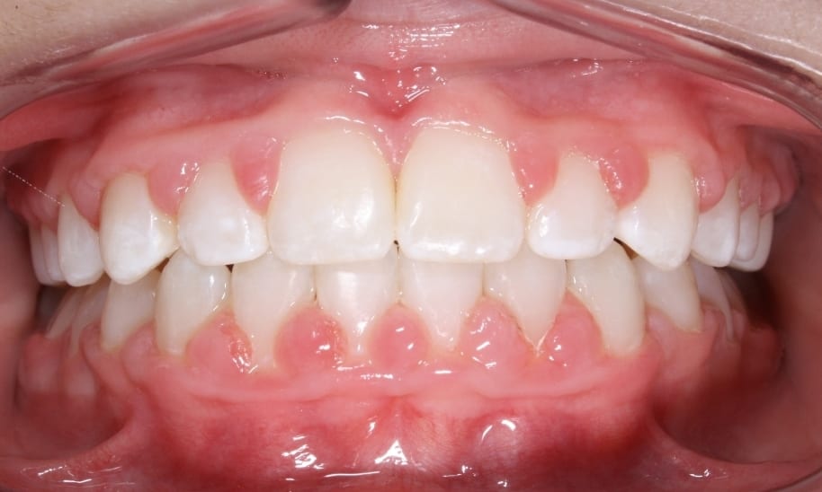
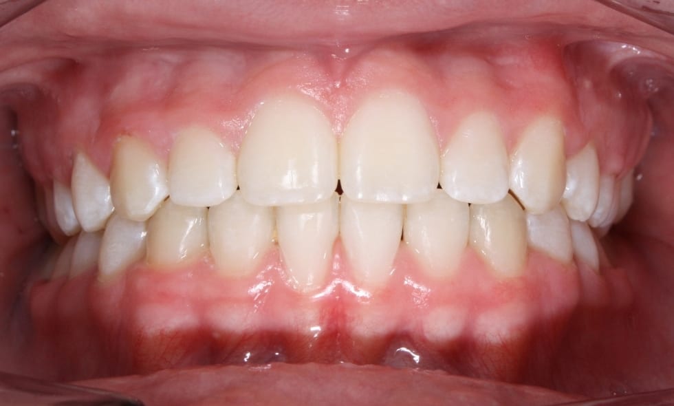
Before and After
See the difference that braces could make for you
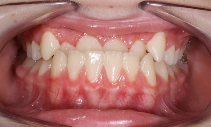
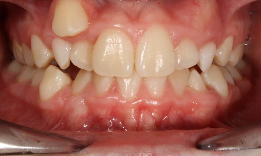
Sam – Before
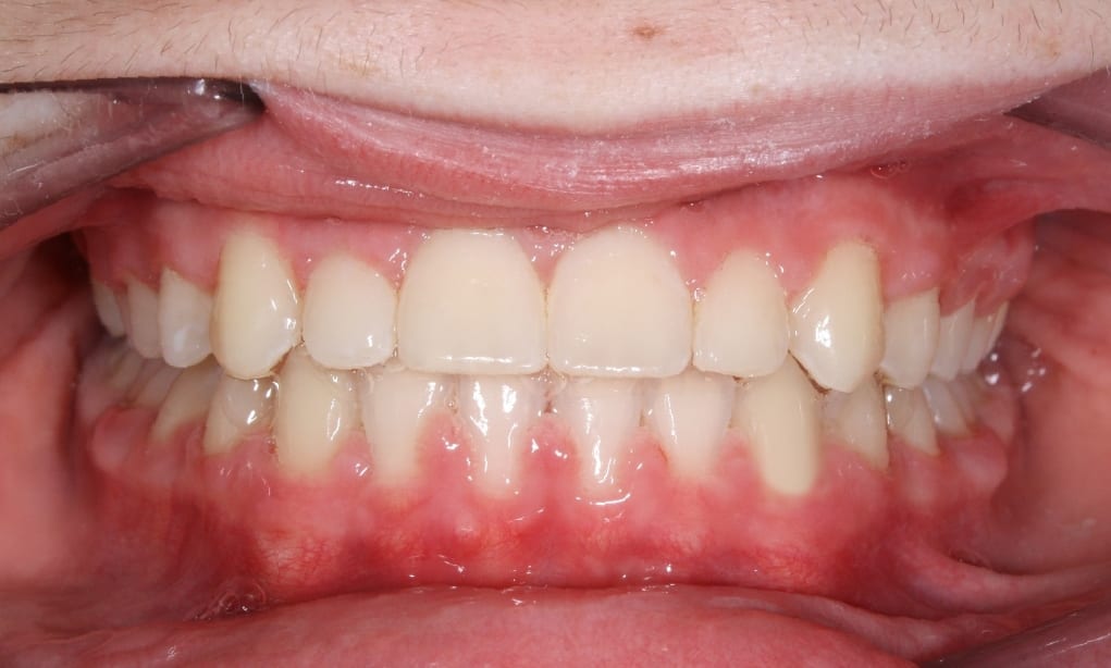
Sam – After
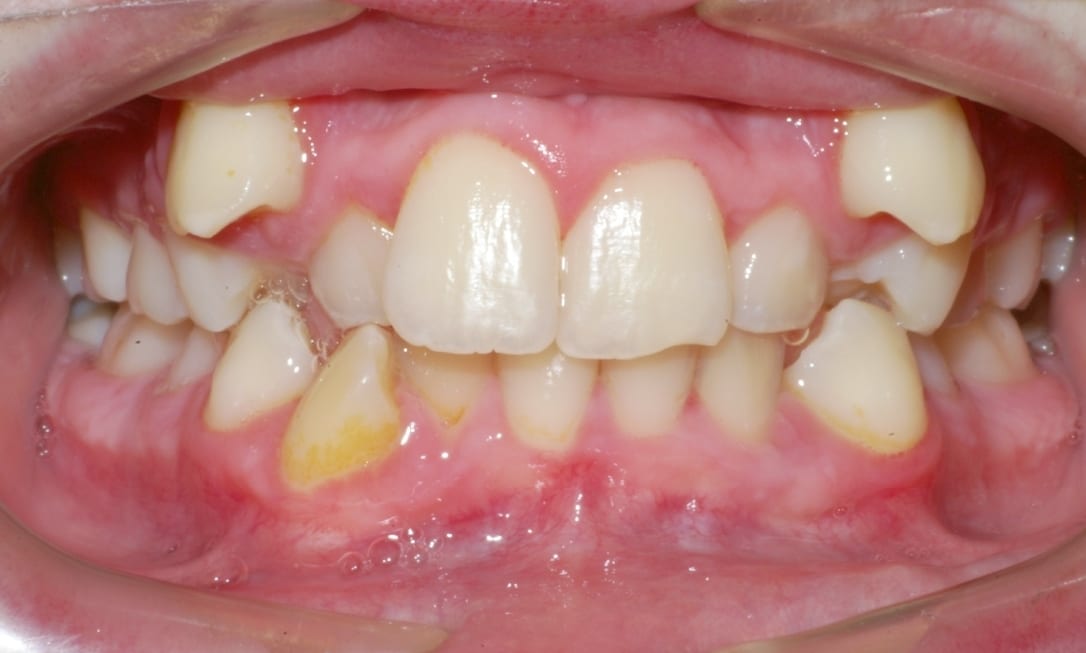
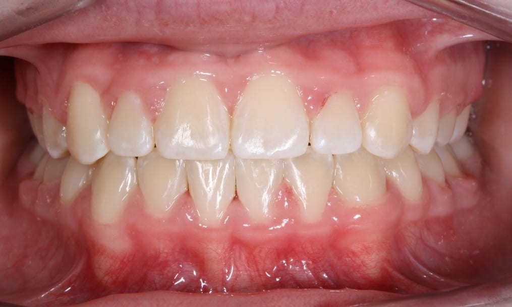
Sebastian – Before
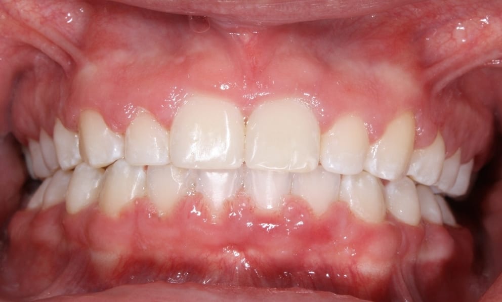
Sebastian – After
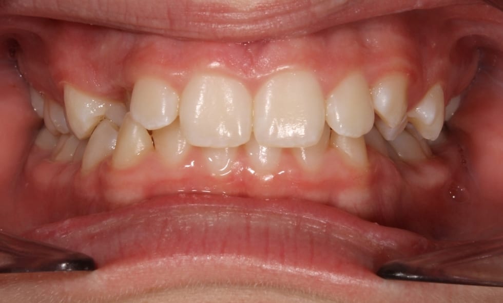
Daniel – Before
Daniel – After
Delany – Before
Delany – After
Rebecca – Before
Rebecca – After
Ali – Before
Ali – After
Bailey – Before
Bailey – After
About
Meet Your Orthodontist
Team
Why McDonough?
Our Lifetime Guarantee
Before and After
Office Tour
Dentofacial Orthopedics
Our Videos
Blog
Patients
FAQ
Your First Visit
Payment Options
Two-Phase Treatment
Children’s Orthodontist
Adult Orthodontics
Choosing an Orthodontist
Services
Braces
Types of Braces
Types of Appliances
Bonding
Clear Aligners
Aligners FAQ
Aligners Videos
Retainers
Dental Monitoring
Contact Us
Request Appointment
Murray Office
Refer A Friend
Refer A Patient (for Doctors)
Careers
(801) 266 2662
Skip to content
Open toolbar
Accessibility Tools
Accessibility Tools
Increase Text
Increase Text
Decrease Text
Decrease Text
Grayscale
Grayscale
High Contrast
High Contrast
Negative Contrast
Negative Contrast
Light Background
Light Background
Links Underline
Links Underline
Readable Font
Readable Font
Reset
Reset